- November 28, 2019
- Adult, Health Articles
Alzheimer’s disease is a common condition that affects the brain. The prevalence of the disease is significantly higher among the elderly population. Alzheimer’s disease has been associated with a large number of adverse effects in the body, including memory loss, poor cognitive function, and, of course, dementia. Those individuals who develop Alzheimer’s disease are also at risk of complications that can lead to death.
What Is Alzheimer’s Disease
Alzheimer’s disease is a condition that is both chronic and progressive. Over time, the disease causes a gradual deterioration of the brain – leading to the destruction of several cognitive functions that a person depends on throughout their lifespan.
While the disease tends to start out slowly, leading to mild signs of dementia, it tends to lead to the development of more serious complications in the long term. This does not only cause difficulty with a person’s ability to think clearly, concentrate on tasks, process information, and utilize their memory function. In addition to these adverse effects, Alzheimer’s disease is also known to cause a deterioration of simple abilities that the person depends on a daily basis – such as washing the dishes, getting dressed, and even taking a bath.
The disease was first recognized by Dr. Alois Alzheimer in the year 1906 when specific brain-related changes were identified in a woman’s brain. The woman had died from a mental disorder that is considered unusual – the patient expressed symptoms that included problems with language, unpredictable behavior, and memory loss. There were physiological changes in the patient’s brain that were not noted in previous brains that the doctor had examined.
Today, Alzheimer’s disease is well-recognized in the medical industry. According to one epidemiology study on Alzheimer’s disease, an estimated 25 million adults worldwide are affected by the condition at the moment. The prevalence of Alzheimer’s disease is also expected to double every two decades – which means as many as 50 million adults may be affected by the year 2040.
Recognizing Alzheimer’s Disease Through Imaging Tests
Imaging tests are often used to assist in the monitoring of a patient’s brain and can sometimes prove useful in the detection of a certain brain-related condition. Even in patients who are suspected of suffering from Alzheimer’s disease, a series of imaging tests can yield an effective way of determining the level of damage that has been dealt with by the disease.
The primary reason Alzheimer’s disease can sometimes be detected with the use of these imaging tests is due to the physiological changes that have been found in the brains of patients suffering from the condition.
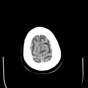
CT Scans
A Computed Tomography, also known as a CT scan, is often utilized to help a physician or specialist rule out other potential causes of cognitive impairment, dementia, and other related symptoms that the patient might experience. Other potential causes of these symptoms may include the development of a tumor in the patient’s brain. Previous episodes of a stroke or subdural hematoma may also cause similar symptoms. These are all conditions that are often identifiable on a CT imaging scan.
Patients suspected of Alzheimer’s disease will likely also undergo a Magnetic Resonance Imaging test, also called an MRI scan. This type of test utilizes a magnetic field, along with a series of radiofrequency pulses. The result is a computerized image of the patient’s head – and, in particular, internal organs that are targeted through the test. In the case of Alzheimer’s disease, the brain will be the target organ – which will then be resonated on a computerized display, based on the findings of the radio frequency pulses combined with the magnetic field used in the test.
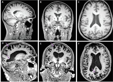
MRI Scans
The use of an MRI test can assist in producing an overview of abnormalities that are present in the patient’s brain. Individuals suspected to have developed Alzheimer’s disease should note that the use of MRI scans can often not produce images that allow the identification of the condition at an early stage, however.
Patients suspected of Alzheimer’s disease will likely also undergo a Magnetic Resonance Imaging test, also called an MRI scan. This type of test utilizes a magnetic field, along with a series of radiofrequency pulses. The result is a computerized image of the patient’s head – and, in particular, internal organs that are targeted through the test. In the case of Alzheimer’s disease, the brain will be the target organ – which will then be resonated on a computerized display, based on the findings of the radio frequency pulses combined with the magnetic field used in the test.
MRI Scans
The use of an MRI test can assist in producing an overview of abnormalities that are present in the patient’s brain. Individuals suspected to have developed Alzheimer’s disease should note that the use of MRI scans can often not produce images that allow the identification of the condition at an early stage, however.
Combined PET/CT Scan
It should be noted that many specialists will combine the use of CT scans and PET scans to provide themselves with a better diagnostic tool for a patient who might be suffering from Alzheimer’s disease.
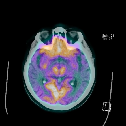
The combination of these scans provides an overview of both the functioning of the brain, along with a view on brain anatomy. The combined images assist in allowing the specialist to identify existing damage that the brain had suffered already while also being able to understand the severity of the disease in terms of cognitive function and the physiological functioning of the brain itself.
PET/MRI Scan
The simultaneous use of PET/MRI allows for the spatial and temporal correlation of the measured signals, thus creating opportunities impossible to achieve through standalone instruments. The importance of PET/MRI scan lies in the fact that various factors such as tau, amyloid, inflammation, perfusion, and metabolic changes influence the cascade of neurodegeneration and functional decline associated with Alzheimer’s disease.
In this case, the PET/MRI scan offers a comprehensive evaluation in a single imaging session and opens new opportunities for the introduction of the two imaging information. Moreover, due to the fact, the atrophy could represent a more downstream event in the course of neurodegeneration, the simultaneous assessment of molecular and functional abnormalities may allow for the more precise staging of Alzheimer’s disease. The use of PET/MRI scans also happens to provide more convenience, especially when elderly and fragile patients undergo the imaging sessions.
Brain-Related Structural Changes Caused By Alzheimer’s Disease
The use of the imaging tests that were described above can provide a more definite view of the structure changes and brain-related damage that a patient had suffered from the presence of Alzheimer’s disease.
In Alzheimer’s patients, the sulci of the brain will generally appear wider, especially as the disease progresses. At the same time, the ridges of the cortex will generally have a thinner appearance. The ventricles, which are cavities found at the center of the patient’s brain, will also start to grow in size gradually.
One of the earliest affected areas of the brain is the hippocampus. In most cases, the disease is diagnosed when the hippocampus is significantly damaged. The hippocampal area is one of the major targets of hallmarks associated with Alzheimer’s disease, such as amyloid plaques, neurofibrillary tangles (NFT), and loss of neurons. The number of NFTs and their spatiotemporal distribution correlate with cognitive decline and progression of Alzheimer’s disease. At the very beginning, NFTs affect the brain in the entorhinal/perirhinal cortex, which impairs the origin of the projection of the perforant pathway to the hippocampus.
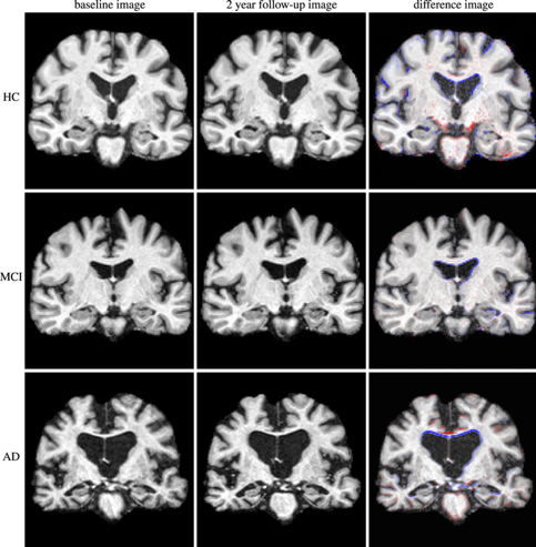
In the hippocampus, NFTs target CA1 first, followed by subiculum, CA2, and CA3. Although NFTs occur alongside the loss of neurons and synapses, it remains unclear whether they are a causative factor in this instance. The spreading of NFTs is also manifested through increased atrophy of the brain, all of which worsens the cognitive decline of a patient. The progressive cerebral atrophy encompasses the atrophy of the hippocampus and is detected by MRI. In most patients, atrophy of the hippocampus, which progresses nonlinearly, is one of the earliest noticeable signs of ongoing neurodegeneration. The most pronounced atrophy associated with Alzheimer’s disease is observed in the anterior hippocampus, which is also a predictive marker of conversion to this disease.
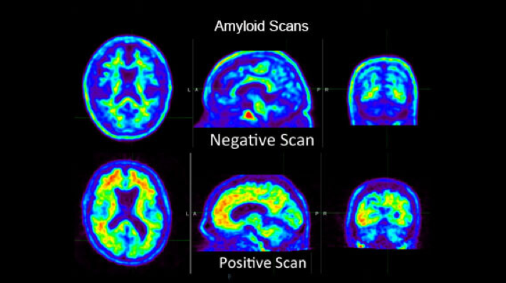
When it comes to brain changes in Alzheimer’s disease, they may begin years or decades before the disease is diagnosed. Although a patient does not experience symptoms in the early stages of the disease, imaging technologies can spot deposits of amyloid-beta. Progression of the disease is observed through cerebral atrophy caused by dendritic and neuronal losses. Besides the hippocampus, the earliest site of atrophy is the entorhinal cortex. The pathologically increased cerebral atrophy starts early and continues relentlessly or at least until patients are severely affected.
Treatment
The standard treatment of Alzheimer’s disease relies on medications such as cholinesterase inhibitors and memantine. In some instances, medications like antidepressants are prescribed to help manage the behavioral symptoms of patients. An important role in the management of Alzheimer’s disease rests upon the education of patients and their families or caregivers. Early referral to a local support group is recommended.
References
https://www.nia.nih.gov/health/what-alzheimers-disease
https://www.ncbi.nlm.nih.gov/pmc/articles/PMC3405821/
https://stanfordhealthcare.org/medical-conditions/brain-and-nerves/dementia/diagnosis/brain-scans.html
https://www.ncbi.nlm.nih.gov/pubmed/29512073
http://jnm.snmjournals.org/content/early/2012/11/09/jnumed.112.105346.full.pdf
https://www.ncbi.nlm.nih.gov/pmc/articles/PMC3978283/
https://www.aafp.org/afp/2011/0615/p1403.html